Medical imaging plays a crucial role in the diagnosis and treatment of various health conditions. Accurate segmentation of the diaphragm from CT images is essential for diagnosing and treating respiratory and related diseases. This article reviews several research papers that propose different approaches for automatic or semi-automatic diaphragm segmentation.
Methodologies
A variety of mathematical and computational methods have been proposed for diaphragm segmentation:
1. 3D active appearance model (2002)
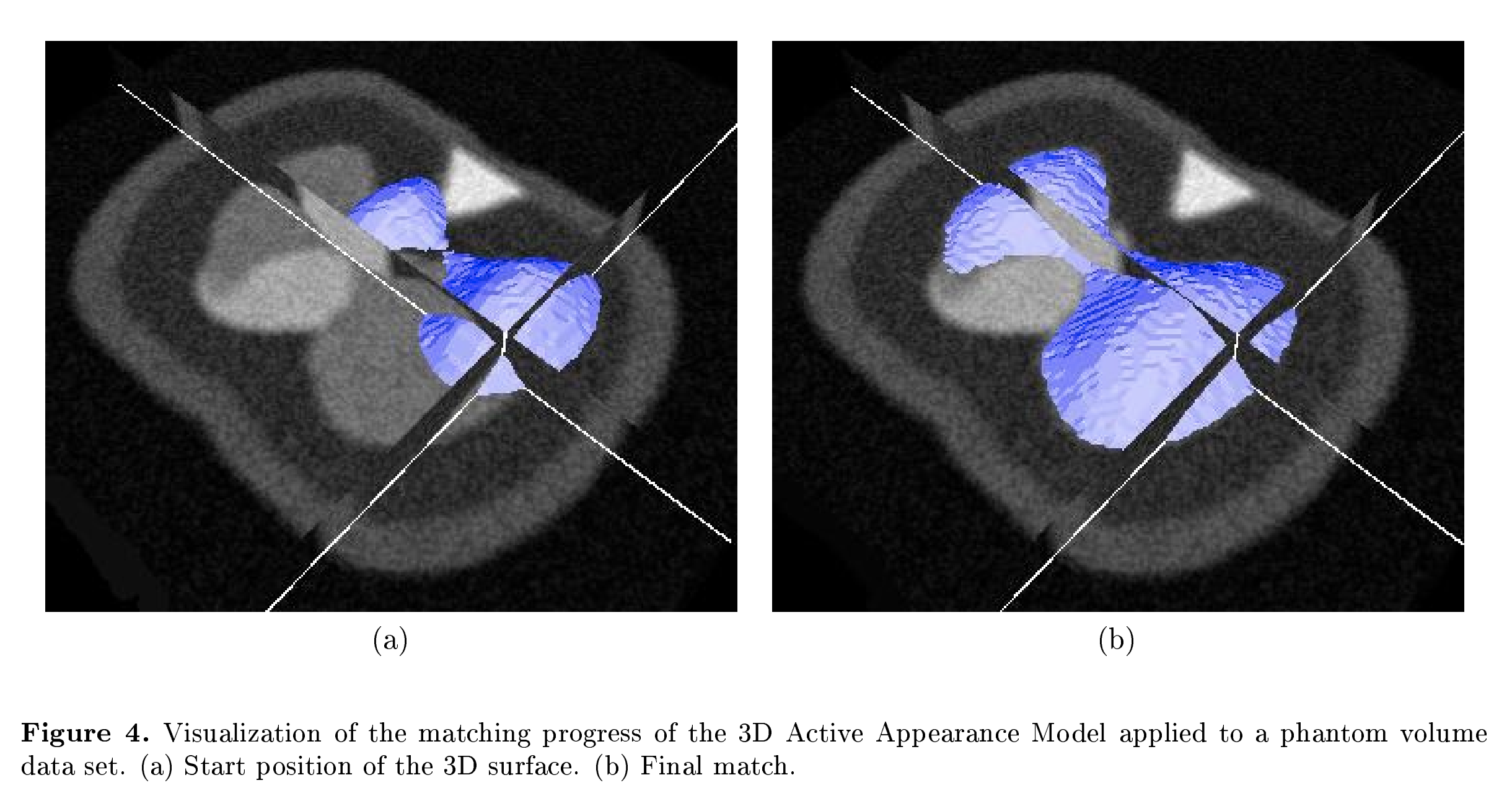
- This mathematical model requires a dataset for training and validation. The heart-diaphragm interface is not considered.
2. Manual modeling using Altair HyperMesh (2006)
3. Automated estimation of the upper surface of the diaphragm in 3-D CT images (2008)
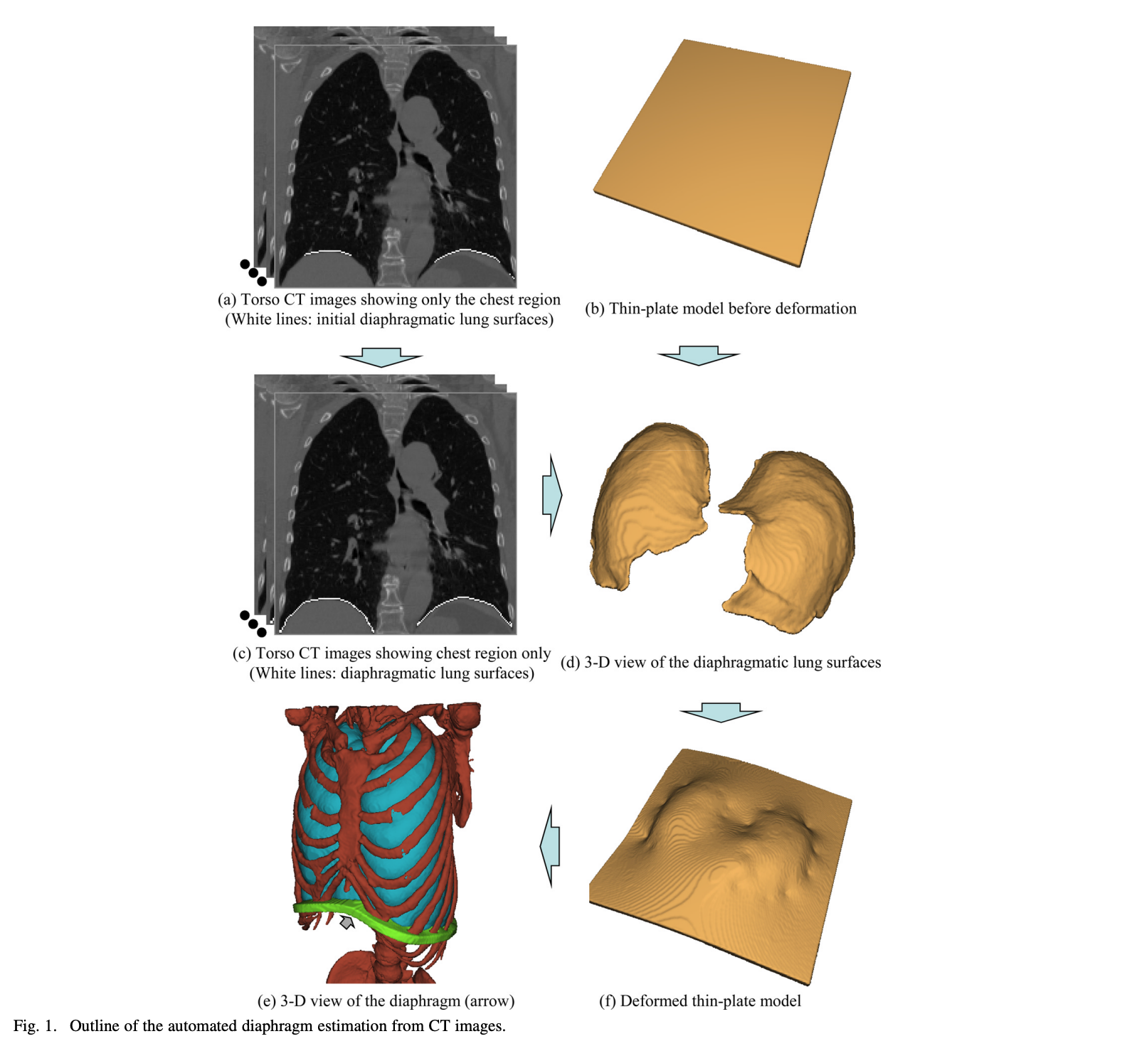
- On each voxel, the normal vector V of the lung surface is calculated. The voxel P on the lung surfaces that satisfies the condition {-(3/4)π < direction of V on P < -(1/4)π} is regarded as the candidate voxel. A thin-plate model is deformed to fit the subsample points on the diaphragmatic lung surface by using a 3D thin-plate spline method.
4. Quadratic surface modeling (2008)
- The shape of the lung is obtained by morphological transformation, and then the lung-diaphragm interface is calculated (no specific elaboration), and finally, the quadratic surface is fitted.
5. Manual segmentation and 3D mesh generation (2008)
- The diaphragm was manually segmented and a 3D mesh generated using Insight SnAP software.
6. Local entropy-based segmentation (2010)
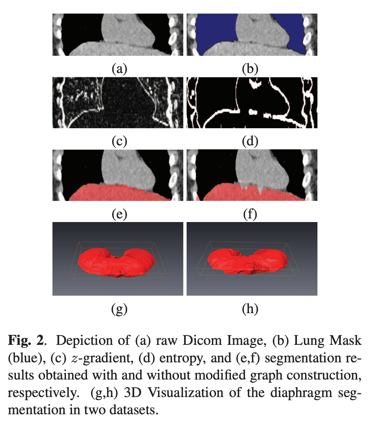
- Does not clarify where the red section stops segmentation.
7. Multiorgan extraction and surface fitting in volumetric CT (2014)
- There is no specific explanation of the 3D ray projection method and no reference to related papers. But Matlab's gridfit surface fitting algorithm is worth trying.
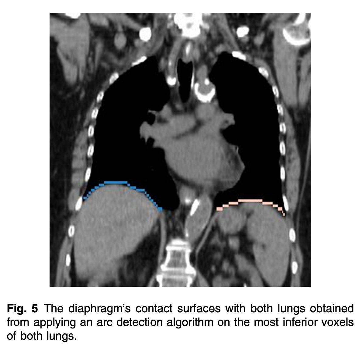
8. Ray projection-based segmentation (2016)
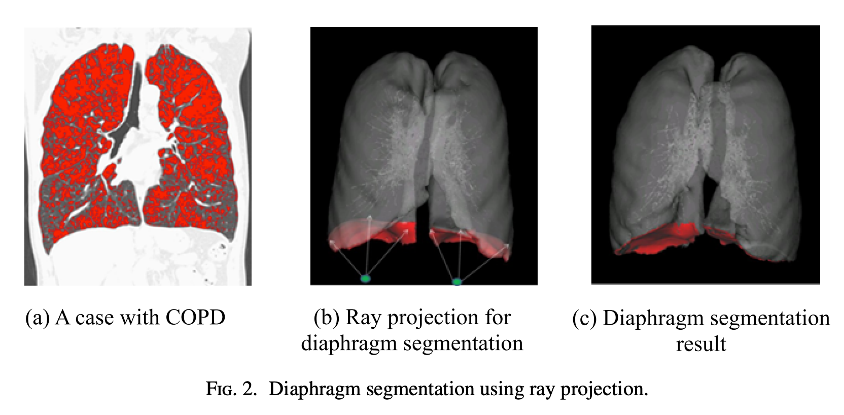
- Refer to Paper 7 (Automatic): 2014-Thoracic cavity segmentation algorithm using multiorgan extraction and surface fitting in volumetric CT
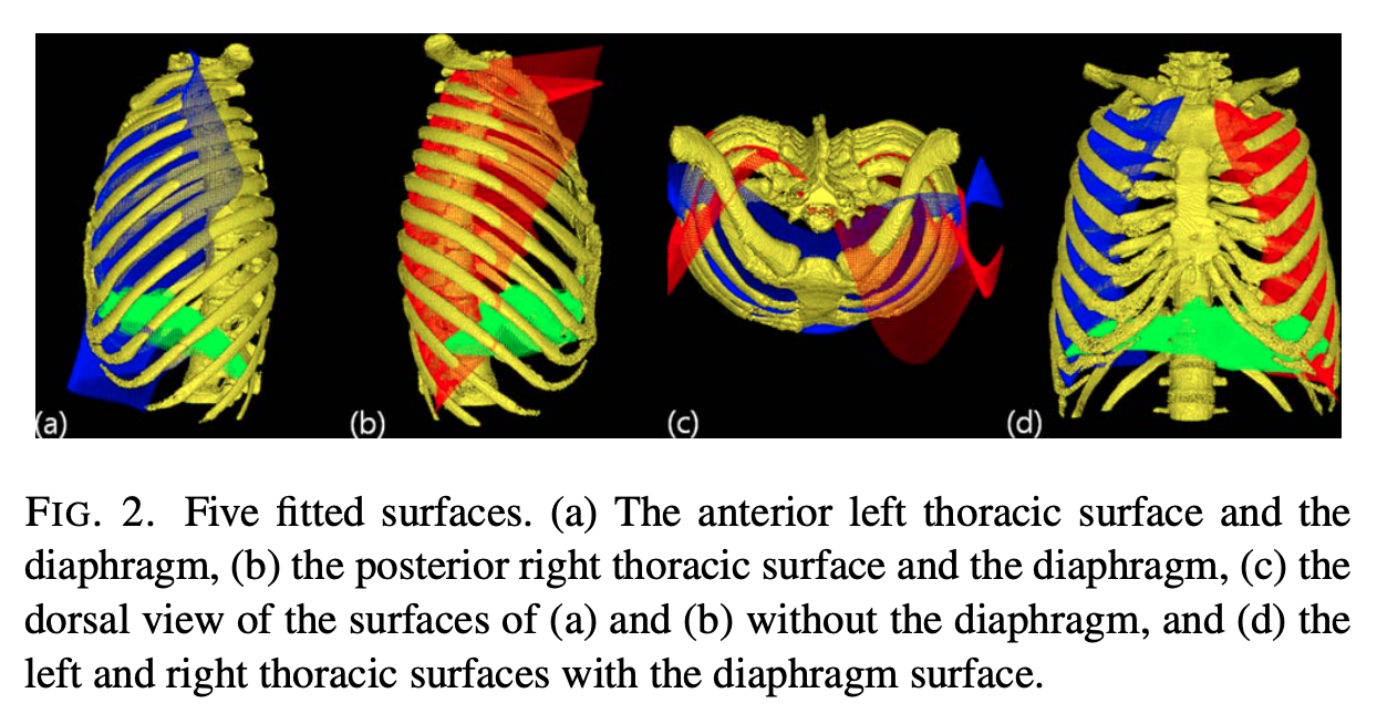
9. Anatomy-based segmentation (2016)
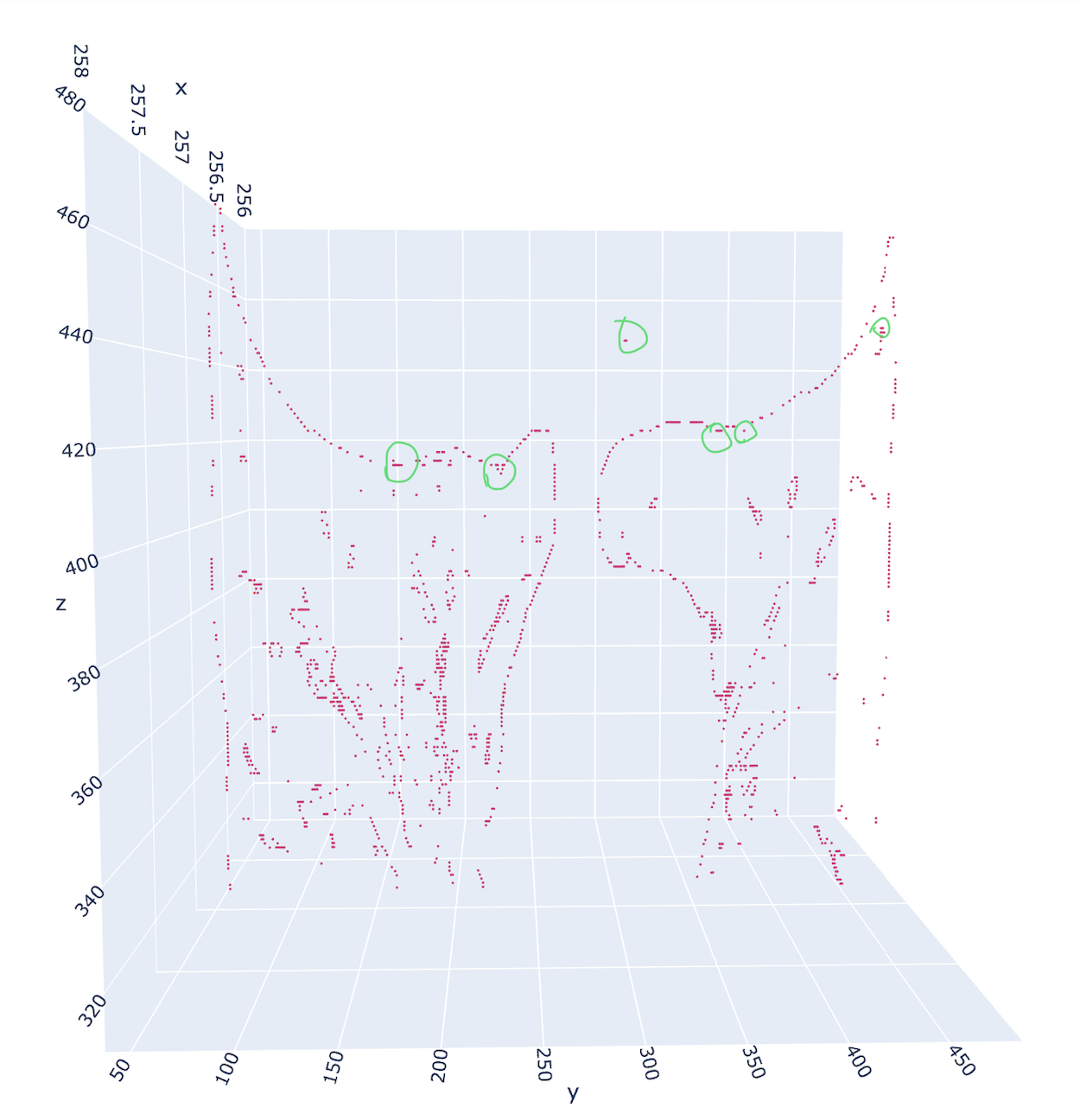
- The algorithm proposed in the paper is unstable. The above figure is a coronal slice arbitrarily selected along the anteroposterior axis (x-axis). If the segment lung-diaphragm interface starts along the horizontal axis (y-axis), the green circles marked in the figure may be misjudged as stops due to gradient change.
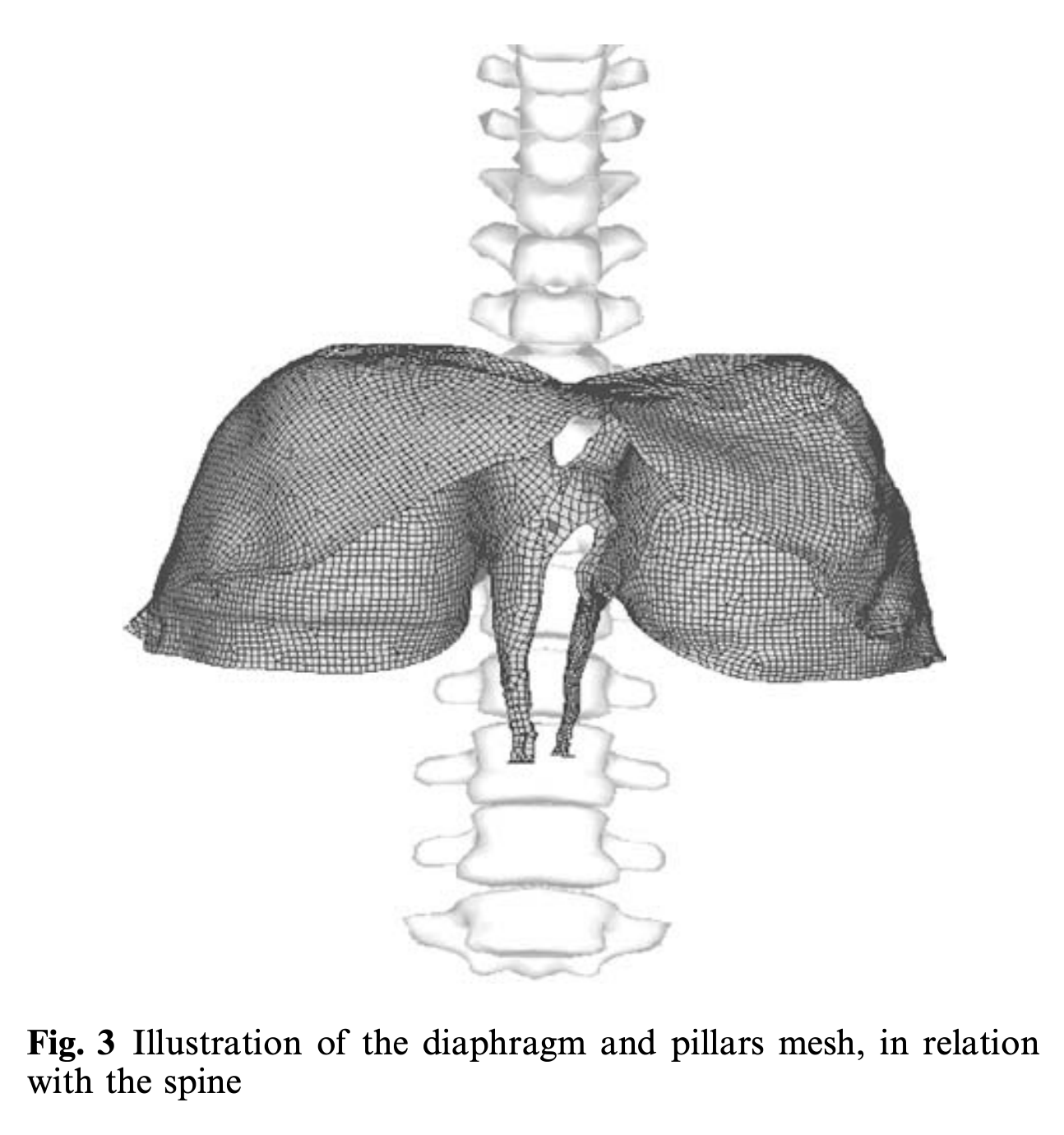
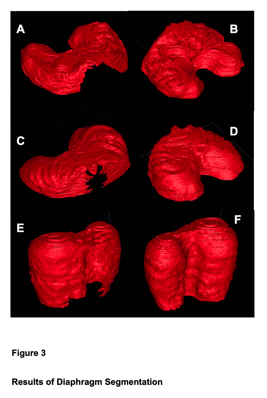
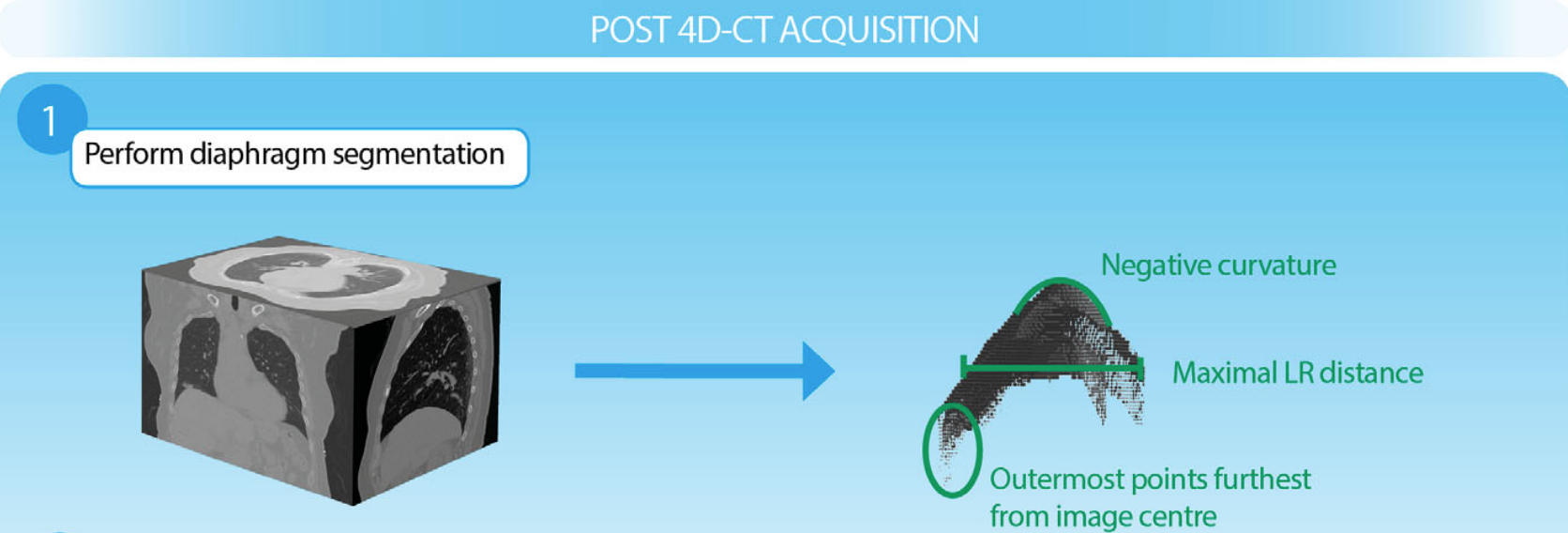
10. Patient-specific diaphragm muscle modeling (2018)
- The development of a patient-specific model of the diaphragm muscle began with modification and use of a previously developed automatic segmentation algorithm for 4DCT images in order to segment the diaphragm volume at end exhalation, which coincides with muscle relaxation. The diaphragm volume was converted into a mesh with hexahedral elements using 3D Slicer.
11. Real-time direct diaphragm tracking (2019)
- It is not clear whether to apply the intensity threshold at the 2D or 3D level; further investigation is required.
Strengths and Weaknesses
Each approach has its own set of advantages and limitations. Manual segmentation, as used in Paper 5, can be time-consuming and requires expert knowledge. Automated methods, such as those proposed in Papers 3, 4, 6, 7, 8, and 9, 11 can be faster and more consistent, but may not be as accurate as manual segmentation.
Methods based on specific features or conditions, such as normal vectors, quadratic surfaces, or local entropy, may perform differently depending on the specific CT images being analyzed. Further investigation is needed to compare their performance in different scenarios.
Conclusion
The development of automated or semi-automated segmentation methods for the diaphragm has important implications for the diagnosis and treatment of respiratory and related diseases. Accurate segmentation can aid in surgical planning, radiation therapy, and assessment of lung function and disease progression.